Chapter 1 — Understanding Fetal Cardiac Orientation & Situs
Before inspecting the four chambers or great arteries, the examiner must reliably determine fetal left from fetal right. Ultrasound screen orientation does not automatically represent true anatomical laterality; screen coordinates change continuously with fetal lie (cephalic vs. breech), fetal spine orientation (anterior, posterior, or lateral), and transducer manipulation.
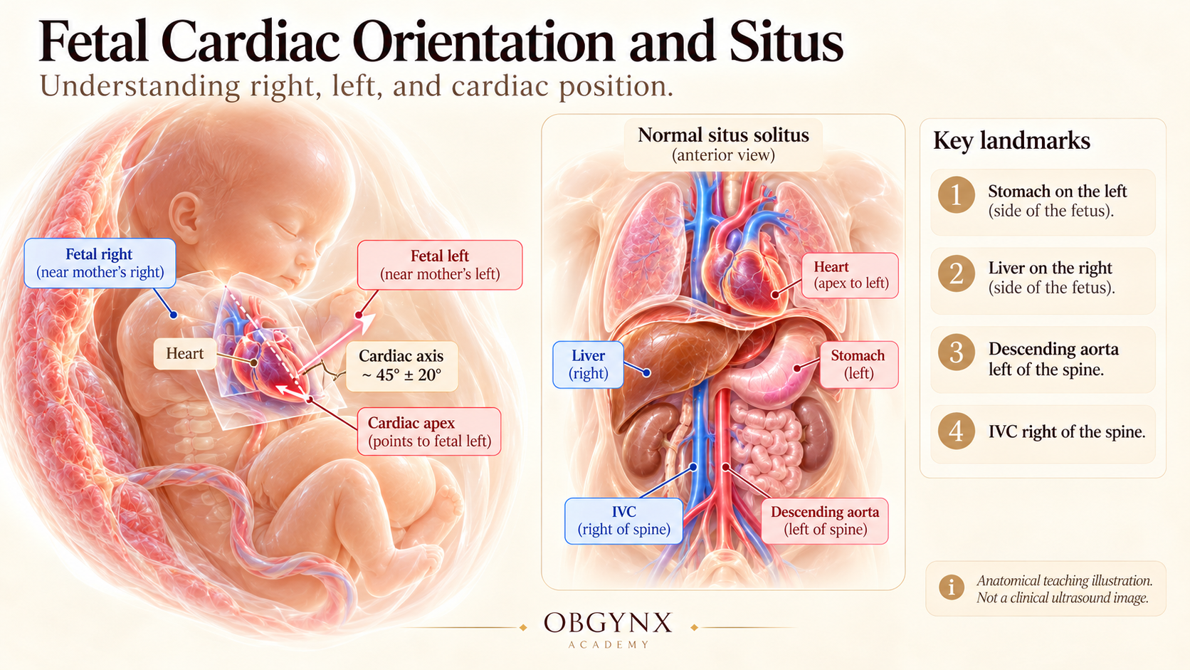
Figure 1: Fetal Cardiac Orientation and Situs — Fetal presentation, identifying true anatomical right and left, leftward cardiac axis (~45° ± 20°), and normal situs solitus relationships.
Identify fetal lie (longitudinal vs transverse) and spine position. In a vertex (cephalic) fetus with spine on the maternal left, the fetal left side is anterior; if spine is on the maternal right, fetal left is posterior. The "right-hand rule" (or mentally placing oneself in the fetal position) confirms anatomical laterality without guesswork.
Situs Solitus: Normal arrangement (stomach, spleen, left atrium, and apex on the left; liver and IVC on the right).Situs Inversus: Mirror-image reversal (incidence of CHD is ~3–5%).Situs Ambiguus (Heterotaxy): Disordered lateralization. Right atrial isomerism (asplenia syndrome) features bilateral right atria, asplenia, midline liver, and severe cyanotic CHD in >90%. Left atrial isomerism (polysplenia syndrome) features bilateral left atria, interrupted IVC with azygos continuation, heart block, and complex CHD.
The normal cardiac axis is 45° ± 20° (range 25°–65°) to the left of the thoracic midline. The heart occupies approximately one-third of the thoracic cross-sectional area, with two-thirds residing in the left hemithorax (levocardia). Deviation of the axis >65° strongly alerts the sonographer to conotruncal CHD, coarctation, or diaphragmatic hernia.
Chapter 2 — The Normal Abdominal Situs View
The first transverse plane of the standard ISUOG sweep is obtained in the upper abdomen just below the diaphragm. Establishing normal abdominal situs is the indispensable prerequisite to evaluating cardiac anatomy.
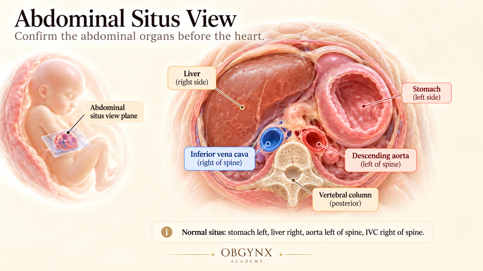
Figure 2: Abdominal Situs View — Transverse cross-section through the upper fetal abdomen demonstrating stomach on the left, liver on the right, descending aorta left of spine, and IVC right of spine.
- Fluid-filled Stomach: Must be located entirely in the left upper quadrant.
- Liver & Gallbladder: Homogeneous liver on the right with teardrop-shaped gallbladder to the right of midline.
- Descending Aorta: Round, pulsatile vessel lying anterior and immediately to the left of the vertebral body.
- Inferior Vena Cava (IVC): Slightly larger, more anterior vessel lying to the right of the midline.
- Intrahepatic Umbilical Vein: Curves gracefully from the anterior abdominal wall towards the right into the portal sinus.
If the aorta and IVC run side-by-side on the same side of the spine (aorta-IVC juxtaposition), right atrial isomerism is practically certain. If the IVC is absent and replaced by a dilated azygos vein posterior to the aorta, left atrial isomerism with interrupted IVC is present.
Chapter 3 — The Normal Four-Chamber View (4CV)
The four-chamber view is the cornerstone of fetal echocardiography. Obtained in an axial thoracic plane showing a complete single rib on each side, it allows rigorous assessment of cardiac size, axis, chamber proportionality, atrioventricular valve offset, and septal integrity.
- Cardiac Size: Occupies approximately 1/3 of the thoracic area (cardiothoracic ratio circumference < 0.50).
- Cardiac Axis: Apex directed to the left at 45° ± 20°.
- Cardiac Position: 2/3 of heart in left hemithorax, 1/3 in right.
- Atrial Symmetry: Two equal-sized atria without intraluminal masses.
- Foramen Ovale Flap: Located in left atrium; flap deflects rhythmically into LA.
- Lower Atrial Septum: Septum primum intact at the cardiac crux.
- Ventricular Symmetry: RV and LV of comparable dimension and wall thickness.
- Interventricular Septum: Intact muscular and membranous septum from apex to crux.
- Two Distinct AV Valves: Both mitral and tricuspid valves open fully and symmetrically.
- AV Valve Offset: Tricuspid septal leaflet inserts 1–2 mm more apical than mitral leaflet.
- Ventricular Morphology: RV contains coarse trabeculae and apical moderator band; LV is smooth-walled with conical apex.
- Pulmonary Veins: At least two veins clearly entering the posterior left atrium.
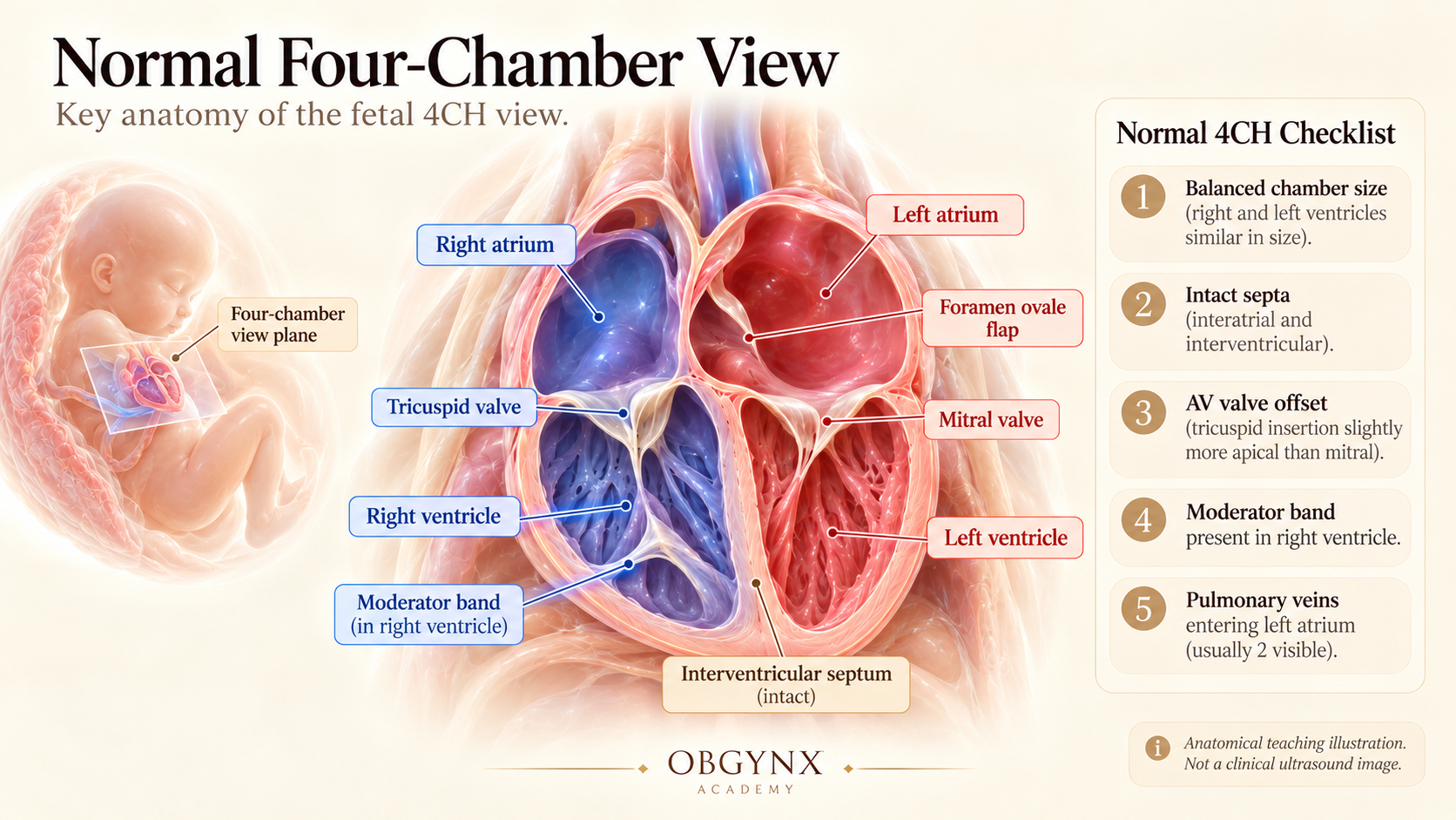
Figure 3: Normal Four-Chamber View (4CV) — Comprehensive anatomical checklist showing balanced ventricular chambers (RV and LV), intact interatrial and interventricular septa, foramen ovale flap in left atrium, pulmonary venous entry, and differential atrioventricular valve offset (tricuspid valve leaflets insert 1–2 mm more apical than mitral valve leaflets).
- Coarse apical muscular trabeculations
- Prominent Moderator Band crossing right ventricular apex
- Septal insertion of tricuspid valve leaflets directly on IVS
- Tripartite anatomy (inlet, trabecular, and infundibulum)
- Smooth septal surface free of chordal attachments
- Conical bullet-shaped apex forming the cardiac apex
- Paired papillary muscles (anterolateral & posteromedial)
- Direct fibrous mitro-aortic continuity (no infundibulum)
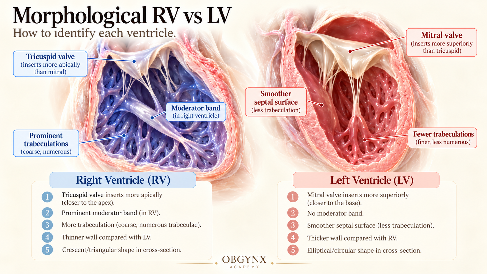
Figure 4: Morphological Ventricular Differentiation — Detailed comparative criteria between the Right Ventricle (RV) with apical moderator band, coarse trabeculae, and apical tricuspid offset, and the Left Ventricle (LV) with smooth septal wall, conical contour, and two distinct papillary muscles.
While a normal 4CV excludes ~50–60% of congenital defects (such as HLHS, AVSD, large muscular VSDs, and Ebstein anomaly), it is entirely blind to conotruncal and aortic arch anomalies. D-TGA, Tetralogy of Fallot, Truncus Arteriosus, Coarctation of the Aorta, and Double Outlet Right Ventricle frequently present with a completely pristine four-chamber view!
Chapter 4 — Left Ventricular Outflow Tract (LVOT)
From the 4CV, angling the transducer slightly cephalad and towards the fetal right shoulder opens the left ventricular outflow tract, demonstrating the ascending aorta exiting the morphological left ventricle.
The cardinal feature of the LVOT view is unbroken septoaortic continuity. The anterior wall of the ascending aorta must merge directly and smoothly with the crest of the muscular interventricular septum. A gap or malalignment indicates a ventricular septal defect with or without an overriding great artery (e.g., Tetralogy of Fallot or Truncus Arteriosus).
The aortic valve leaflets should be thin and mobile, opening fully to the vessel walls during systole without thickening or doming. Color Doppler reveals a smooth, laminar blue jet crossing the valve without turbulent aliasing (peak systolic velocity normally 60–90 cm/s).
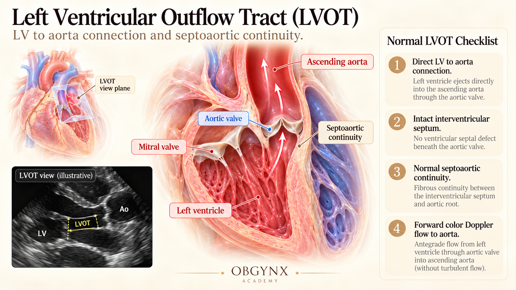
Figure 5: Left Ventricular Outflow Tract (LVOT) — Uninterrupted septoaortic continuity with thin, mobile aortic valve leaflets, absence of muscular subaortic conus, and direct origin of the ascending aorta from the morphological left ventricle.
Chapter 5 — Right Ventricular Outflow Tract (RVOT)
Sweeping further cephalad from the LVOT brings the right ventricular outflow tract into view, originating from the anterior, heavily trabeculated morphological right ventricle.
In normal human anatomy, the two great arteries cross each other perpendicularly. The Main Pulmonary Artery arises anteriorly and crosses in front of the ascending aorta towards the left shoulder. If both great arteries emerge parallel to each other in the same plane, transposition of the great arteries (d-TGA) is present.
The main pulmonary artery wraps around the ascending aorta and promptly bifurcates into the right and left branch pulmonary arteries (RPA and LPA). Documenting this bifurcation confirms that the vessel is the true pulmonary trunk and not a solitary truncus or misplaced aorta.
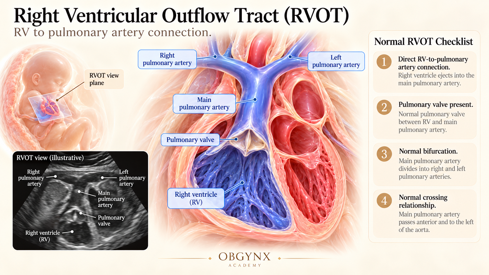
Figure 6: Right Ventricular Outflow Tract (RVOT) — Demonstration of normal perpendicular great artery crossover: the anterior Main Pulmonary Artery originates from the RV, wraps across the ascending aorta, and promptly bifurcates into right and left branch pulmonary arteries.
Chapter 6 — The Three-Vessel View (3VV)
The three-vessel view is an axial plane across the upper mediastinum demonstrating the cross-sectional profiles of the main pulmonary artery, ascending aorta, and superior vena cava in a clean diagonal sequence.
From anterior-left to posterior-right, the vessels must adhere strictly to the rule: Main Pulmonary Artery > Ascending Aorta > Superior Vena Cava. Any reversal (e.g., aorta significantly larger than pulmonary artery) suggests pulmonary stenosis, Tetralogy of Fallot, or hypoplastic right heart. Conversely, a diminutive aorta suggests critical aortic coarctation or aortic stenosis.
Finding only 2 vessels suggests Truncus Arteriosus, Pulmonary Atresia, or Aortic Atresia. Finding 4 vessels indicates a Persistent Left Superior Vena Cava (PLSVC) located to the extreme left of the pulmonary artery.
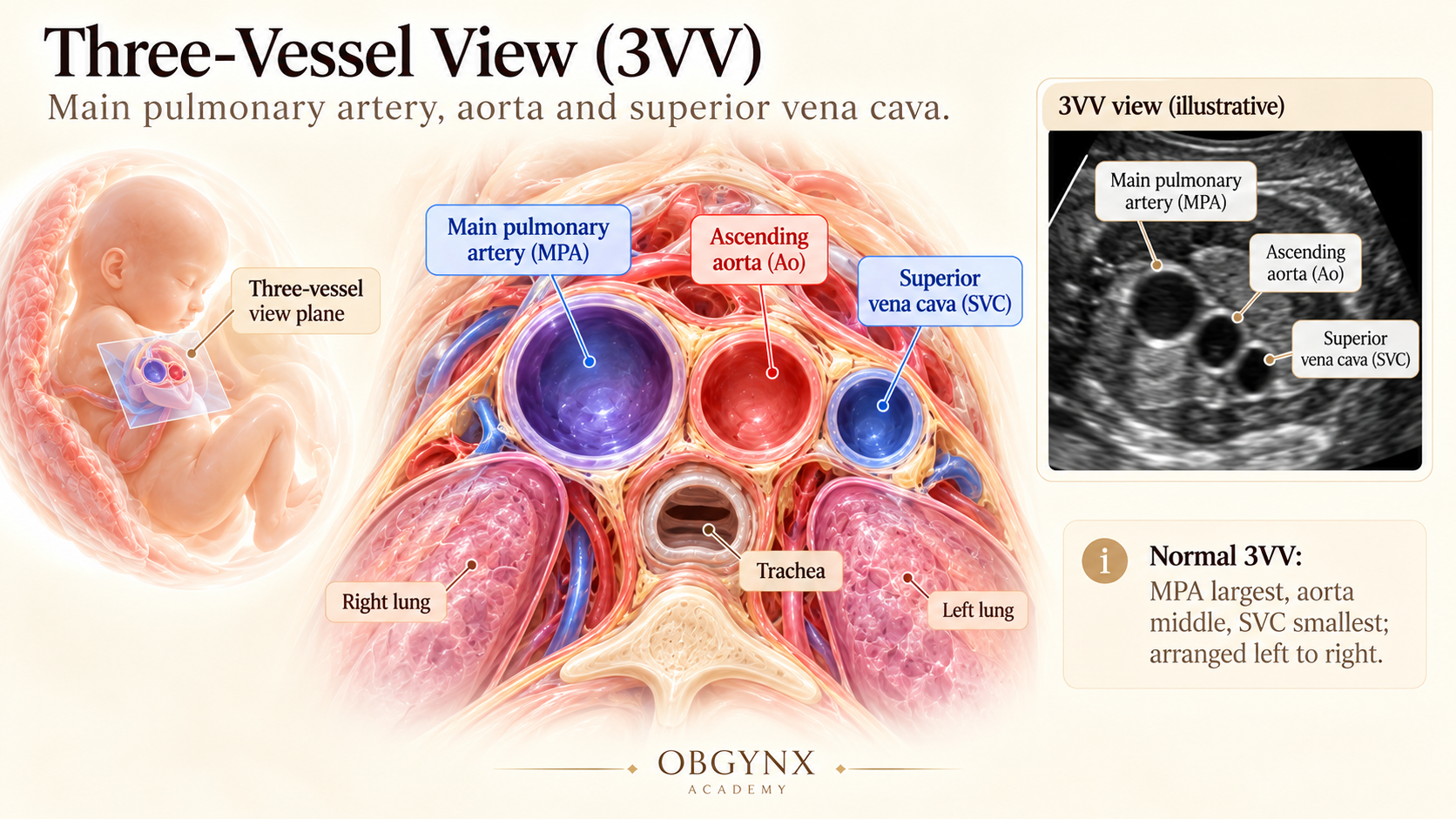
Figure 7: Three-Vessel View (3VV) — Upper mediastinal axial plane demonstrating strict spatial ordering and caliber hierarchy from anterior-left to posterior-right: Main Pulmonary Artery (PA) > Ascending Aorta (Ao) > Superior Vena Cava (SVC) aligned in a straight oblique line.
Chapter 7 — The Three-Vessel-and-Trachea View (3VT / 3VTV)
The 3VT view is arguably the single most high-yield plane for detecting aortic arch anomalies and ductal-dependent lesions. Obtained slightly higher in the upper mediastinum than the 3VV, it showcases both the transverse aortic arch and ductal arch meeting at the descending aorta.
The ductal arch (left limb) and aortic arch (right limb) converge posteriorly into the descending thoracic aorta, forming a sharp 'V'. Crucially, the apex and limbs of this 'V' must lie entirely to the LEFT of the echogenic ring of the trachea.
In a Right Aortic Arch, the aortic limb runs to the right of the trachea, forming a 'U-sign' with the left ductus arteriosus that encircles the trachea and esophagus. In a Double Aortic Arch, both right and left arches persist, creating a complete vascular ring.
On color Doppler, blood flow in both the aortic arch and ductal arch must travel in the same forward direction (concordant color, typically blue). Reverse flow in the ductal arch indicates ductal-dependent pulmonary circulation (e.g. pulmonary atresia); reverse flow in the aortic arch indicates critical coarctation or aortic atresia.
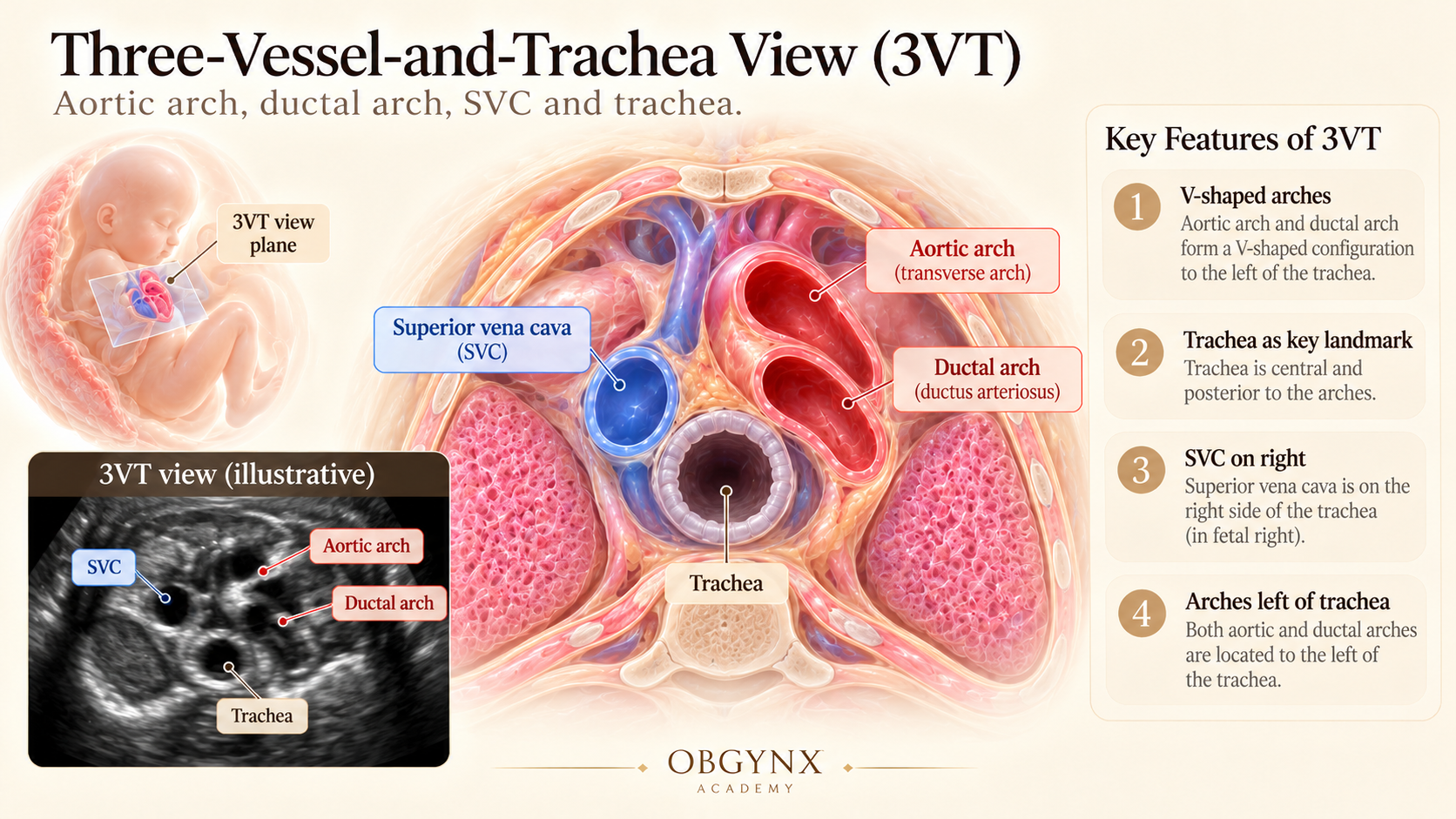
Figure 8: Three-Vessel-and-Trachea View (3VTV) — Convergence of the ductal arch and transverse aortic arch forming an acute left-sided 'V-sign' meeting at the descending aorta, positioned strictly to the left of the echogenic trachea with concordant forward color Doppler flow.
Chapter 8 — Aortic Arch vs Ductal Arch: Sagittal Differentiation
When transverse imaging suggests an arch anomaly, sagittal parasagittal sweeps allow distinct differentiation of the aortic arch from the ductal arch based on shape, origin, and branching pattern.
- Origin: Arises from deep within the center of the thorax from the LV.
- Contour: Tight, high-curvature hairpin arch ("candy cane").
- Head & Neck Branches: Gives rise to three distinct branches from its convexity: brachiocephalic artery, left common carotid artery, and left subclavian artery.
- Pathology: Coarctation typically occurs at the isthmus just distal to the left subclavian insertion.
- Origin: Arises immediately anteriorly from the right ventricle.
- Contour: Flat, wide-angle curve without sharp bending ("hockey stick").
- No Branching: Completely unbranched conduit running directly into the descending thoracic aorta.
- Function: Transports ~90% of RV stroke volume directly to the systemic circulation.
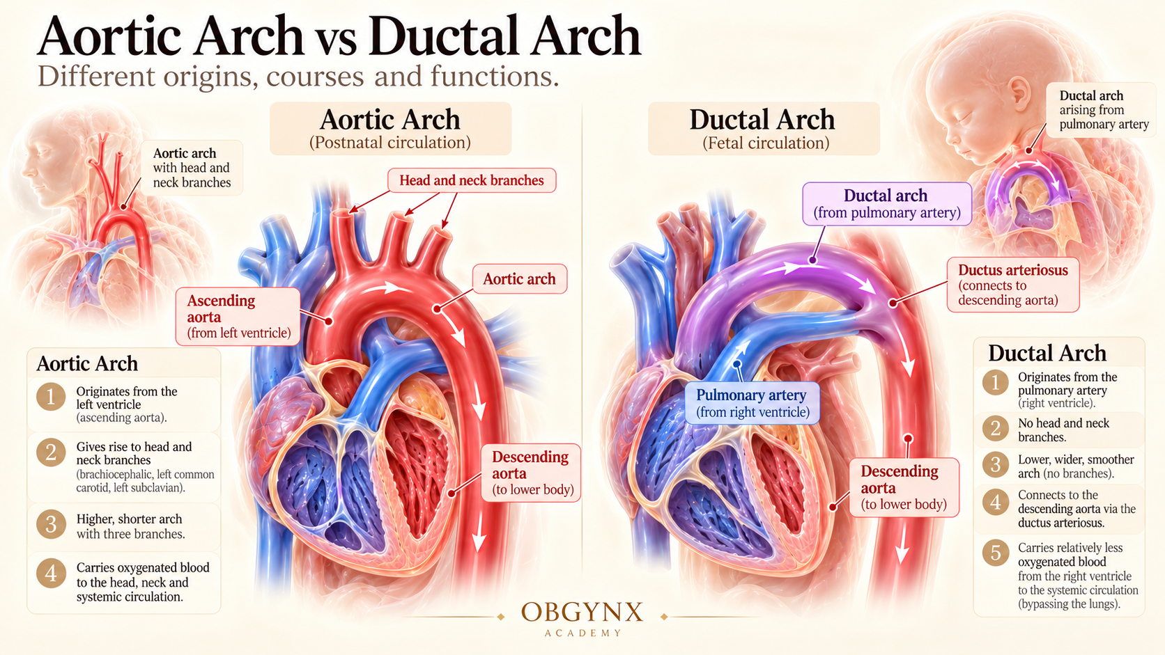
Figure 9: Sagittal Arch Differentiation — Parasagittal sonographic view contrasting the tight hairpin curvature and brachiocephalic head-and-neck branches of the Aortic Arch ('Candy Cane') with the anterior, wide-curved, unbranched Ductal Arch ('Hockey Stick') connecting into the descending thoracic aorta.
Chapter 9 — Additional Specialized Fetal Echo Views
While the five transverse planes constitute the mandatory universal screening examination, a comprehensive specialist fetal echocardiogram incorporates specialized planes and Doppler interrogations:
Demonstrates the SVC entering superiorly and the IVC entering inferiorly into the morphological right atrium. Essential for confirming normal systemic venous return and ruling out interrupted IVC.
Low-velocity color Doppler (< 20 cm/s) and pulsed Doppler waveforms documenting pulsatile biphasic forward flow into the left atrium to exclude Total Anomalous Pulmonary Venous Return (TAPVR).
Shows the circular left ventricle ("doughnut") surrounded anteriorly by the crescent-shaped right ventricle ("croissant"). Assesses regional wall motion and interventricular septal flattening.
Peak systolic velocities across aortic (< 100 cm/s) and pulmonary (< 90 cm/s) valves with insonation angle < 30°, ruling out critical semilunar valve stenosis or regurgitation.
Chapter 10 — The Five Transverse Scanning Planes: The ISUOG Master Protocol
The International Society of Ultrasound in Obstetrics and Gynecology (ISUOG) guidelines establish that a sequential cephalad sweep through five transverse planes constitutes the highest sensitivity protocol for prenatal cardiac anomaly detection.
| Plane | Level | Probe Motion | Critical Check |
|---|---|---|---|
| 1. Upper Abdomen | Subdiaphragmatic | Transverse cross-section | Stomach left, liver right, aorta left of spine, IVC right of aorta. |
| 2. Four-Chamber (4CV) | Mid-thorax | Slide 2–4 mm cephalad | 1:1 chamber balance, 45° axis, crux offset, moderator band, intact septum. |
| 3. LVOT | High mid-thorax | Tilt towards right shoulder | Aorta emerges from LV, continuous with IVS without overriding. |
| 4. RVOT / 3VV | High thorax | Tilt towards left shoulder | MPA crosses anterior to aorta, bifurcates into LPA/RPA; PA > Ao > SVC. |
| 5. 3VT | Superior mediastinum | Slide slightly cephalad | Aortic & ductal arches meet as acute 'V' strictly left of trachea. |
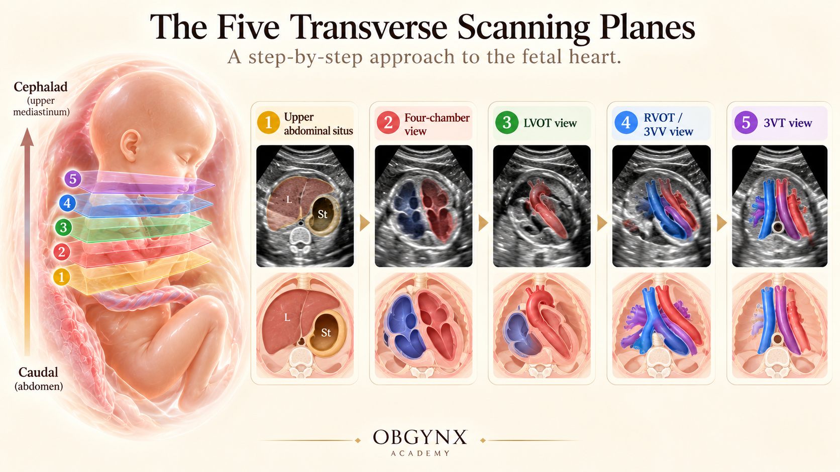
Figure 10: The Five Transverse Scanning Planes — Comprehensive visual guide depicting the sequential ISUOG fetal screening protocol: Plane 1 (Abdomen/Situs), Plane 2 (Four-Chamber View), Plane 3 (LVOT), Plane 4 (RVOT & 3VV), and Plane 5 (Three-Vessel-and-Trachea View).
Chapter 11 — Normal vs Abnormal Pattern Recognition Matrix
Structured side-by-side diagnostic comparative reference across the entire fetal echocardiographic sweep:
| View | Normal Finding | Abnormal Finding | Key Differentials | Next Step |
|---|---|---|---|---|
| Situs | Stomach left, liver right | Stomach right, midline liver | Situs Inversus, Heterotaxy | Assess IVC/Aorta position & spleen |
| 4CV Size | Heart = 1/3 thorax area | Cardiomegaly (>1/2 thorax) | Ebstein, severe TR/MR, anemia | Evaluate for hydrops & arrhythmias |
| 4CV Offset | TV offset 1–2 mm apical | Linear insertion of valves | AVSD (Trisomy 21 association) | Fetal karyotype / microarray |
| LVOT | Septoaortic continuity | Overriding aorta > 50% | Tetralogy of Fallot, Truncus | Inspect pulmonary valve & 22q11 deletion |
| RVOT | Arteries cross perpendicularly | Arteries emerge parallel | D-TGA (Transposition) | Plan delivery at cardiac surgery center |
| 3VT V-Sign | V-apex left of trachea | U-sign around trachea | Right Aortic Arch, Vascular Ring | Check aberrant subclavian & 22q11 |
Chapter 12 — Common Scanning Mistakes & Technical Pitfalls
Even experienced sonographers encounter common technical and interpretive traps. Here is how to diagnose and correct each:
The Trap: Tangential imaging that cuts through the ventricular apex prematurely, underestimating ventricular length and creating artificial chamber disproportion.
The Fix: Ensure ribs on both sides of the thorax are symmetrical and single before measuring chamber areas.
The Trap: Insonating parallel to the interventricular septum (apical 4CV) causes pseudoloss of signal in the thin perimembranous septum, falsely diagnosing a VSD.
The Fix: Rotate the probe to insonate the septum perpendicularly (subcostal 4CV) and confirm with color Doppler (PRF 30–40 cm/s).
The Trap: In the third trimester (34–38 weeks), physiological right ventricular dominance is normal (RV:LV ratio up to 1.3:1). Diagnosing coarctation on chamber disproportion alone causes excessive false positives.
The Fix: Check the 3VT view and sagittal aortic arch. Coarctation requires transverse arch hypoplasia, an isthmic shelf, and abnormal flow.
The Trap: Concluding the examination after obtaining a reassuring four-chamber view. D-TGA has a 100% normal 4CV in almost all cases.
The Fix: Mandatory execution of the cephalad sweep to document arterial crossover in every single fetus without exception.
Chapter 13 — Clinical Diagnostic Takeaway: The Master Algorithm
From Normal Anatomy to Abnormal Pattern Recognition: A 5-step clinical algorithm for daily practice.