Clinical Educational Notice — DRAFT (Medical Review Required)
This scientific masterclass is intended exclusively for certified ultrasound practitioners, maternal-fetal medicine subspecialists, pediatric cardiologists, and OBGYN trainees. All fetal diagnostic algorithms, biometric cutoff values, and intervention criteria must be correlated with official ISUOG / ASE clinical practice guidelines and institutional fetal cardiology consultation.
1. Recognizing a Small Left Ventricle: Observation vs Diagnosis
When a sonographer encounters a four-chamber view where the left ventricle appears smaller than the right, the single most critical clinical realization is this: a small left ventricle is an observation, never a standalone diagnosis. It represents the anatomical starting point of a multidisciplinary diagnostic fork that encompasses benign gestational variation, discrete valvar or arch obstruction, evolving myocardial remodeling, and catastrophic single-ventricle malformations.
In normal fetal mid-gestation (18 to 22 weeks), the left and right ventricular chambers demonstrate close geometric symmetry, with an internal diameter ratio approaching approximately 1:1. However, as pregnancy advances into the late third trimester (beyond 30 to 32 weeks), physiological right ventricular dominance normally emerges, producing a mild physiological disproportion where the right ventricle can be up to 15% to 20% larger than the left ventricle. Recognizing this gestational progression is critical to preventing false-positive referrals for Hypoplastic Left Heart Syndrome (HLHS) or coarctation.
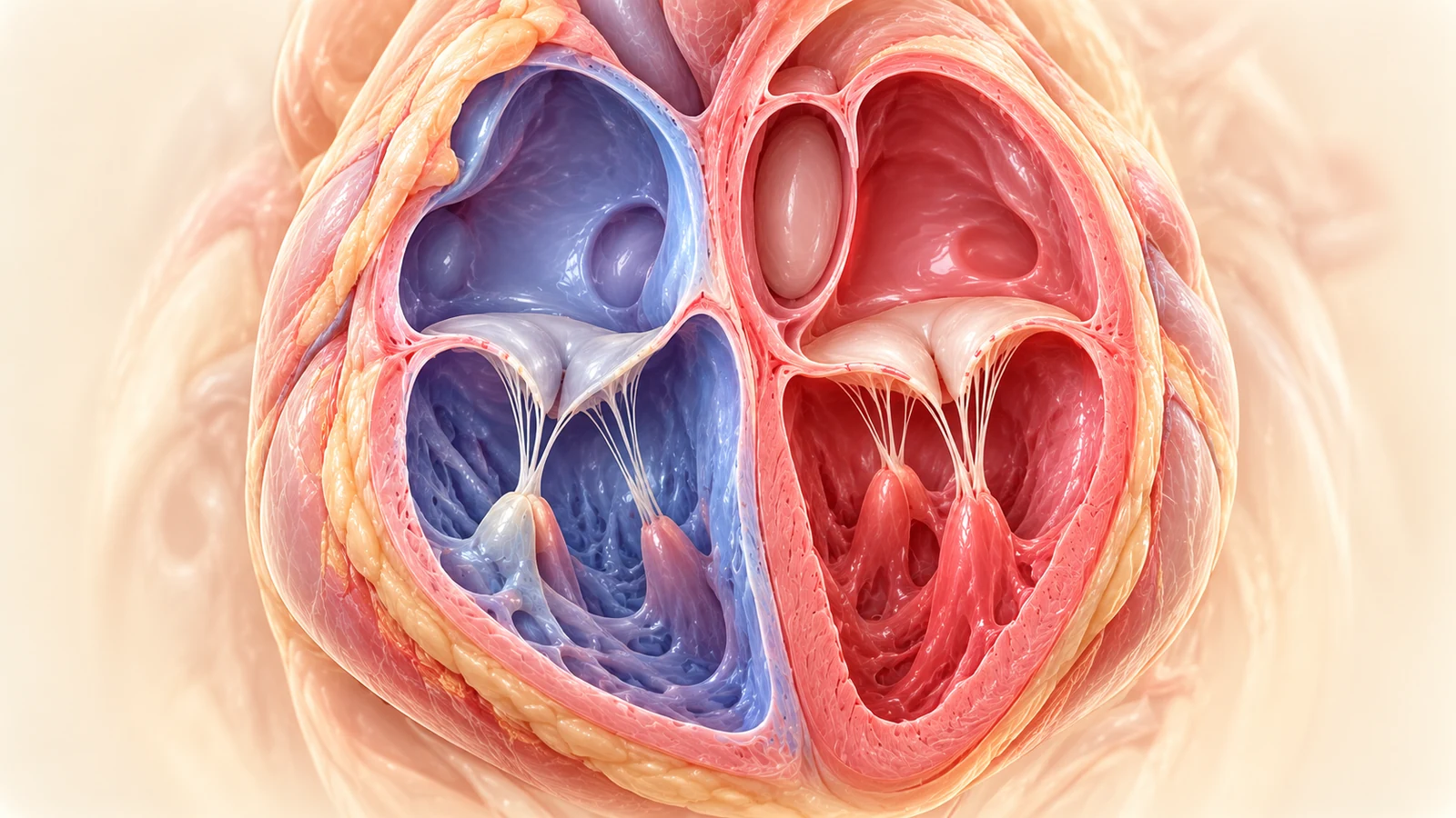 Click to zoom
Normal Four-Chamber Anatomy: Ventricular Symmetry & Apex Formation
High-resolution anatomical section through the fetal thorax demonstrating normal ventricular symmetry in mid-gestation. The smooth-walled, bullet-shaped morphological left ventricle forms the true cardiac apex, while the trabeculated right ventricle with moderator band lies directly beneath the anterior chest wall. Normal tricuspid-mitral valve offset is preserved at the cardiac crux.
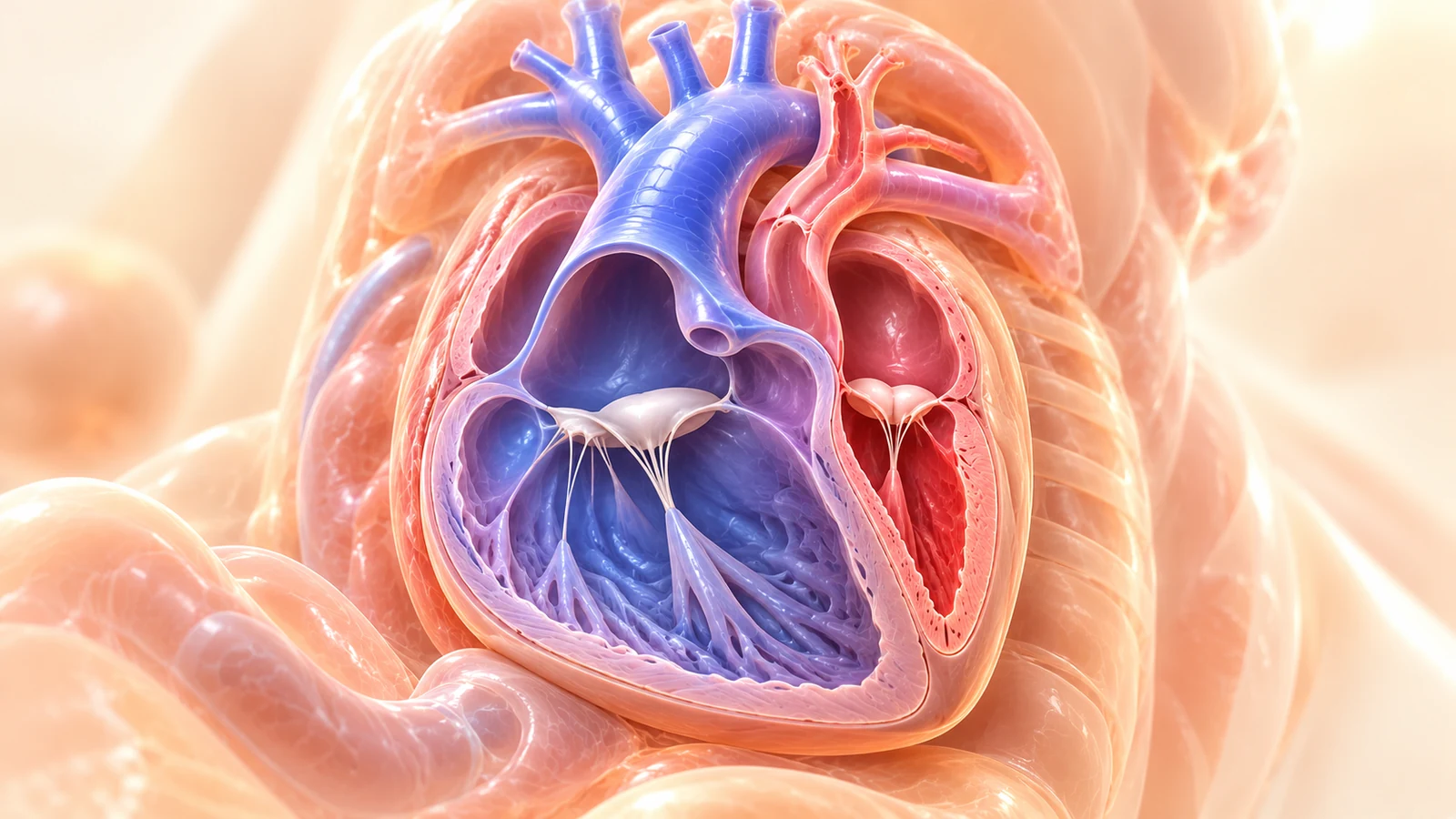 Click to zoom
Normal vs Small Left Ventricle: Visual Disproportion Spectrum
Side-by-side comparative atlas showing normal ventricular proportions (left) versus true left ventricular hypoplasia (right). In the abnormal heart, the apex is formed exclusively by the enlarged, hypertrophied right ventricle, while the diminutive left ventricle is recessed and fails to reach the apex. Note the narrow mitral orifice and diminutive left atrium.
Four Mandatory Rules Before Confirming Ventricular Disproportion:
2. The Master Small Left Heart Diagnostic Fork
When the fetal left ventricle appears small, do not jump to a single entity. Systematically test the eight anatomical checkpoints below. Change the parameters to explore how the clinical differential, pathophysiology, risk tier, and recommended next ultrasound views adapt in real time.
Diagnostic Fork Simulator: Left Ventricle Appears Small
Suspected Fetal Coarctation of the Aorta (Ventricular & Arterial Disproportion)
Hemodynamic Mechanism: Right-dominant ventricular disproportion (RV > LV) and great artery disproportion (PA > Ao) with relative narrowing of the distal transverse arch and aortic isthmus.
- LV is smaller than RV but retains normal apical morphology, biphasic mitral inflow, and antegrade aortic flow
- Aortic isthmus z-score < -2 with bidirectional flow or diastolic forward flow tapering
- Frequently accompanied by persistent left SVC or bicuspid aortic valve
- Important uncertainty: prenatal findings are predictive but NOT definitively diagnostic until postnatal ductal closure
- Sagittal aortic arch view with isthmus caliber and Doppler
- 3VV / 3VT caliber ratio measurements
- Targeted look for persistent left SVC (coronary sinus dilation) and VSD
The Master Small Left Heart Diagnostic Fork Decision Matrix
Open the full high-resolution 8-branch clinical decision matrix in medical lightbox.
3. Hypoplastic Left Heart Syndrome (HLHS): The Anatomical Spectrum
Hypoplastic Left Heart Syndrome (HLHS) accounts for approximately 2% to 3% of all congenital cardiac malformations and represents the archetypal severe ductal-dependent left-sided obstructive lesion. In HLHS, the morphological left ventricle, mitral valve, aortic valve, and ascending aorta are underdeveloped to a degree incapable of supporting systemic circulation.
Crucially, HLHS is not a single uniform anatomical entity. It encompasses a spectrum of phenotypic variants characterized by varying combinations of mitral and aortic valve atresia versus stenosis:
MA / AA: The Slit-Like Cavity
Complete absence of mitral inflow. The left ventricle is tiny, slit-like, or completely microscopic, recessed within the posterior ventricular wall. The ascending aorta is thread-like (1-2 mm) and carries pure retrograde flow for the coronary arteries.
MS / AA: Hypertensive & Globular
Blood enters through a pin-point mitral valve but cannot escape through the atretic aortic root. This generates severe intracavitary hypertension, prominent endocardial fibroelastosis, severe mitral regurgitation, and a thick-walled globular cavity.
MS / AS: The Borderzone LV
Both valves are hypoplastic and stenotic. Left ventricular dimensions are borderline. Determining whether this heart can sustain a biventricular repair or requires a single-ventricle palliation is one of the greatest challenges in fetal cardiology.
Fetal & Neonatal Hemodynamics of Classic HLHS:
In the fetus with HLHS, in-utero survival is normally preserved because the right ventricle performs virtually the entire systemic cardiac output. Blood returning to the right atrium is pumped through the pulmonary artery and traverses the wide ductus arteriosus into the descending aorta to supply the lower body and placenta. Crucially, the upper body (head, neck vessels) and the coronary arteries are perfused by retrograde flow traveling backward down the transverse aortic arch and ascending aorta.
The Neonatal Crisis: At birth, as pulmonary vascular resistance drops and the ductus arteriosus begins physiological closure, systemic perfusion rapidly fails, leading to profound cardiogenic shock, metabolic acidosis, and death unless continuous intravenous Prostaglandin E1 (Alprostadil) is promptly initiated.
4. Critical Fetal Aortic Stenosis & The Evolving HLHS Timeline
Critical fetal aortic stenosis represents one of the few true emergencies in fetal cardiology where in-utero intervention may alter the developmental natural history of the heart. The lesion is defined by severe obstruction of the aortic valve leaflets resulting in marked left ventricular afterload, elevated intracavitary pressure, and impaired systolic ejection.
A critical pitfall is assuming that a small left ventricle is always small from conception. In many cases of critical AS, the left ventricle is initially dilated, spherical, and hypertensive in early mid-gestation. Over weeks, severe subendocardial ischemia induces dense collagen deposition known as Endocardial Fibroelastosis (EFE), rendering the myocardium stiff and non-compliant, ultimately arresting cavity growth and causing it to remodel into an end-stage hypoplastic chamber.
The 4 Stages of Evolving HLHS in Critical Aortic Stenosis
Stage 1: Severe Outflow Obstruction with Dilated Globular LV (18–22 Weeks)
Aortic valve leaflets are thickened with restricted excursion and a high-velocity turbulent jet (>2.0 m/s). The LV is dilated, spherical, and hyperdynamic. Antegrade flow in the ascending aorta is preserved, and foramen ovale shunting remains physiological (right-to-left).
Fetal aortic balloon valvuloplasty is a highly specialized catheter intervention performed under ultrasound guidance in designated tertiary centers. Candidate selection is complex and requires balanced multi-parameter scoring (adequate LV long-axis length, preserved LV pressure generation, and physiological mitral inflow). The procedure carries recognized procedural risks (fetal bradycardia, hemopericardium, intrauterine fetal demise) and does not guarantee biventricular circulation; candidates must be evaluated by multidisciplinary fetal cardiac boards rather than simplified scoring calculators.
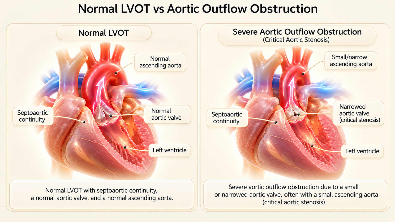 Click to zoom
Normal LVOT vs Critical Fetal Aortic Stenosis
Side-by-side comparative illustration: Normal LVOT with smooth antegrade laminar ejection (left) versus Critical Aortic Stenosis (right). In critical AS, the aortic valve leaflets are thickened, hyperechoic, and immobile. The left ventricle is severely dilated, globular, and hyper-trabeculated with bright endocardial fibroelastosis, ejecting a turbulent high-velocity jet.
Click to zoomEvolving HLHS: Ventricular Remodeling and Function Cascade
The pathophysiological cascade of evolving HLHS. In mid-gestation, severe aortic stenosis causes left ventricular pressure overload and spherical dilatation. Over subsequent weeks, progressive endocardial fibroelastosis, subendocardial ischemia, and mitral inflow restriction arrest myocardial growth, transforming the dilated ventricle into an end-stage hypoplastic cavity.
5. Suspected Fetal Coarctation of the Aorta: Pitfalls & Reality
Coarctation of the aorta (CoA) remains one of the most difficult prenatal cardiovascular diagnoses in all of fetal medicine. It is notorious for generating both significant false-positive rates (unnecessary parental distress and intervention) and false-negative rates (neonatal collapse following unexpected ductal closure at home).
The root reason for this diagnostic difficulty lies in normal fetal hemodynamics. In utero, the fetal right ventricle normally handles approximately 60% of combined ventricular output, with the vast majority flowing across the ductus arteriosus directly into the descending thoracic aorta. Only roughly 10% of total cardiac output traverses the aortic isthmus (the narrow segment between the left subclavian artery and the ductal insertion). Consequently, the fetal isthmus is naturally the narrowest zone of the aortic arch even in healthy fetuses.
Furthermore, the discrete coarctation shelf is a localized infolding of the aortic media located juxtaductally. As long as the ductus arteriosus is widely patent, blood flows effortlessly past this area. The shelf only protrudes into the aortic lumen to create significant pressure gradient after the ductus arteriosus begins physiological constriction in the first 24 to 72 hours of postnatal life.
Multi-Parameter Coarctation Suspicion Matrix
Check the findings observed in your ultrasound examination. Note: Guidelines emphasize that no single biometric ratio is independently pathognomonic; confidence increases when multiple concordant markers align.
Even with high-grade multi-parameter suspicion, up to 40% to 50% of prenatally suspected coarctation cases demonstrate normal arch calibers postnatally without requiring intervention. Conversely, false-negative cases occur. Therefore, antenatal counseling must emphasize planned delivery at a cardiac center with active PGE1 readiness and serial neonatal echocardiography, rather than promising a definitive prenatal confirmation.
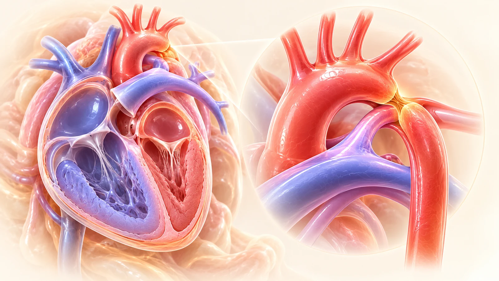 Click to zoom
Normal Aortic Arch vs Coarctation Suspicion
High-resolution sagittal view of the fetal aortic arch. Normal smooth 'candy-cane' arch (left) versus suspected coarctation (right). Note the discrete narrowing at the aortic isthmus (between the left subclavian artery and the ductal insertion) and the prominent right ventricular and pulmonary arterial dominance.
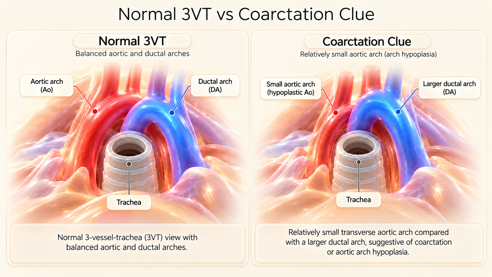 Click to zoom
Normal vs Hypoplastic Transverse Aortic Arch in 3VT View
Three-Vessel and Trachea (3VT) comparative plane: Normal V-configuration with roughly equal arch calibers (left) versus Transverse Arch Hypoplasia (right). The aortic arch is noticeably smaller than the dilated ductal arch, producing marked caliber discrepancy to the left of the trachea.
6. Aortic Arch Hypoplasia & Interrupted Aortic Arch (IAA)
A clear distinction must be maintained between four related yet anatomically distinct entities:
- Aortic Isthmus Hypoplasia: Localized narrowing restricted to the segment between the left subclavian artery and the ductal insertion.
- Diffuse Transverse Arch Hypoplasia: Uniform underdevelopment of the entire arch extending between the innominate artery and the ductal junction.
- Discrete Coarctation: A localized posterolateral shelf of arterial tissue projecting into the lumen at the juxtaductal site.
- Interrupted Aortic Arch (IAA): Complete anatomic luminal discontinuation between the ascending and descending thoracic aorta.
Interrupted Aortic Arch is subclassified based on the Celoria and Patton anatomical classification:
- Type A (30% to 40%): Interruption occurs distal to the origin of the left subclavian artery (isthmic interruption).
- Type B (50% to 60%): Interruption occurs between the left common carotid artery and the left subclavian artery. Critical note: Type B IAA carries a greater than 50% association with 22q11.2 microdeletion (DiGeorge syndrome) and is almost universally accompanied by a posterior malalignment ventricular septal defect with subaortic stenosis.
- Type C (<5%): Interruption occurs between the innominate and left common carotid arteries.
Click to zoomAortic Isthmus and Ductal Arch Anatomical Relationship
Detailed anatomical diagram of the juxtaductal region in the fetal thorax. Demonstrates the physiological relationship between the aortic isthmus (low-flow segment) and the high-flow ductus arteriosus, explaining why the coarctation shelf remains hidden until ductal closure.
Click to zoomNormal vs Interrupted Aortic Arch (IAA) Subtypes
Anatomical schematic and fetal ultrasound correlation of Interrupted Aortic Arch. Illustrates Type A (interruption distal to left subclavian), Type B (interruption between left carotid and left subclavian, strongly linked to 22q11.2 deletion), and Type C (interruption between innominate and left carotid arteries).
7. Unbalanced Atrioventricular Septal Defect (AVSD)
Whenever a small left ventricle is identified, the examiner must immediately scrutinize the cardiac crux. In typical HLHS or coarctation, the crux demonstrates two separate atrioventricular valves with preserved differential offset (tricuspid inserting more apically than mitral).
In an Atrioventricular Septal Defect (AVSD), there is failure of the superior and inferior endocardial cushions to fuse, creating a single common atrioventricular orifice with a common AV valve, absence of the normal offset, a primum ASD, and an inlet VSD.
In approximately 10% to 15% of complete AVSD cases, the common valve does not divide symmetrically across the ventricular septum; instead, it is committed preferentially to one ventricular cavity. When committed predominantly to the right ventricle, the left ventricle receives insufficient inflow and becomes severely hypoplastic—termed Right-Dominant Unbalanced AVSD.
Recognizing an unbalanced AVSD transforms clinical counseling: this is not an isolated left ventricular defect, but a complex cushion anomaly frequently linked to heterotaxy syndromes (right or left isomerism) or Trisomy 21, requiring a single-ventricle palliative surgical track rather than a simple coarctation or biventricular repair.
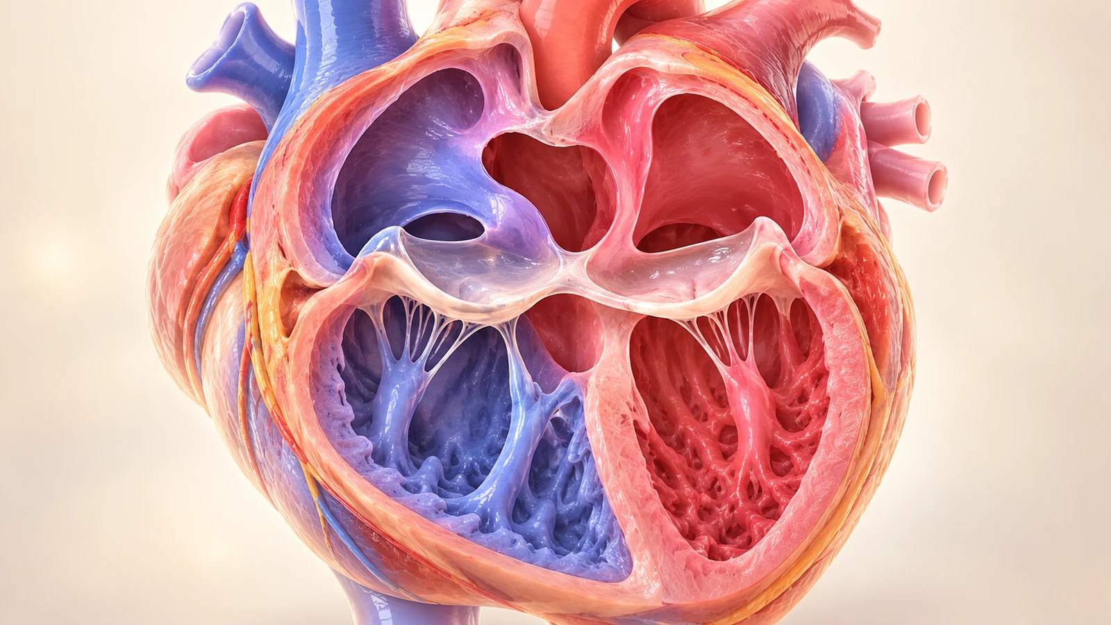 Click to zoom
Normal Crux vs Right-Dominant Unbalanced AVSD
Crux comparison: Normal differential AV valve offset (left) versus Unbalanced Atrioventricular Septal Defect (right). In right-dominant AVSD, a single common AV valve sits across an absent central cardiac crux, with >60% of the valve orifice opening into the right ventricle, leaving the left ventricle under-filled and hypoplastic.
8. Other Causes of a Relatively Small Left Ventricle
Beyond classic HLHS, critical AS, coarctation, and unbalanced AVSD, multiple distinct hemodynamics can alter ventricular loading and produce an apparent or genuine small left ventricle:
A PLSVC draining into the coronary sinus dramatically enlarges the coronary sinus along the posterior left atrioventricular groove. This physically encroaches upon the left atrium and mitral annulus, redirecting inflow and causing secondary relative under-filling of the left ventricle.
When all four pulmonary veins fail to connect to the left atrium and instead drain into the systemic venous circulation (e.g., supracardiac innominate vein or infracardiac ductus venosus), the left atrium receives zero direct pulmonary venous return, leading to reduced left-sided volume.
Premature constriction or anatomical redundancy/aneurysm of the septum primum with restricted right-to-left interatrial shunting limits the primary supply of oxygenated blood to the left ventricle, leading to secondary chamber hypoplasia and severe fetal distress.
In severe early-onset FGR, extreme placental vascular resistance elevates right ventricular afterload while brain-sparing vasodilation alters cerebral and isthmic resistance, often amplifying visual chamber asymmetry without an intrinsic structural cardiac defect.
9. The Mitral Valve Diagnostic Fork: Annulus, Leaflets & Inflow
The mitral valve is the gateway to left ventricular cavity growth. A normal fetal mitral valve consists of anterior and posterior leaflets suspended by chordae tendineae to two distinct anterolateral and posteromedial papillary muscles.
When interrogating the mitral valve, the examiner must evaluate three distinct parameters:
- Annular Diameter: Measured in maximum diastole from hinge point to hinge point, indexed to gestational age z-scores (normal z > -2).
- Leaflet Mobility & Morphology: Thin, pliable leaflets opening fully versus thickened, echogenic, doming leaflets (stenosis) or an imperforate fibrous diaphragm (atresia).
- Spectral Doppler Inflow Profile: Normal fetal inflow demonstrates distinct early passive diastolic filling (E-wave) and late atrial active contraction filling (A-wave), where A is normally greater than E (physiological compliance pattern).
Click to zoomNormal vs Abnormal Mitral Valve Inflow Doppler Profiles
Pulsed Doppler waveforms across the mitral valve: Normal biphasic E and A wave filling (left), monophasic high-velocity restrictive filling in severe stenosis (center), and flatline absent flow in complete mitral atresia (right).
10. The Aortic Valve Diagnostic Fork: Antegrade vs Retrograde
Tracing the left ventricular outflow tract (LVOT) into the aortic root is the decisive step for distinguishing between valvar atresia, critical stenosis, and downstream arch lesions:
Trileaflet or bicuspid valve opening briskly during systole; peak velocity 70–100 cm/s; smooth laminar Doppler envelope.
Thickened, doming, dysplastic cusps; high-velocity systolic jet (>200 cm/s); prolonged ejection time; post-stenotic dilation.
Imperforate fibrous plate; absent antegrade flow; root measures 1–2 mm; color Doppler shows pure retrograde perfusion from ductus.
Click to zoomNormal vs Abnormal Aortic Valve Flow Velocities
Spectral Doppler interrogation of the aortic root: Normal smooth laminar peak velocity ~70-100 cm/s (left), turbulent jet >200 cm/s in Critical Aortic Stenosis with post-stenotic envelope (center), and absent/retrograde flow in Aortic Atresia (right).
11. Aortic Arch & 3VV/3VT Assessment: Calibers & Direction
The 3-Vessel and Trachea (3VT) view is arguably the highest-yield screening plane for detecting aortic arch hypoplasia and ductal-dependent circulation. In this axial plane through the upper fetal mediastinum:
- The ductal arch (arising from the pulmonary trunk) and the transverse aortic arch converge to the left of the trachea in an acute 'V-shape'.
- Both arches normally exhibit roughly comparable calibers (ductal arch slightly larger, but transverse arch easily identifiable).
- Color Doppler confirms that blood in both the aortic arch and ductal arch flows in the same direction (antegrade, away from the transducer, encoded blue).
In severe left heart obstruction (HLHS, severe aortic coarctation with isthmus hypoplasia, aortic atresia), two major anomalies become immediately evident:
- Caliber Discrepancy: The transverse arch is diminutive, thread-like, or invisible alongside a massively dilated ductal arch.
- Retrograde Flow (Ductal Steal): Color Doppler reveals discordant color coding: the ductal arch flows blue (antegrade), while the aortic arch flows red (retrograde toward the neck and coronary vessels).
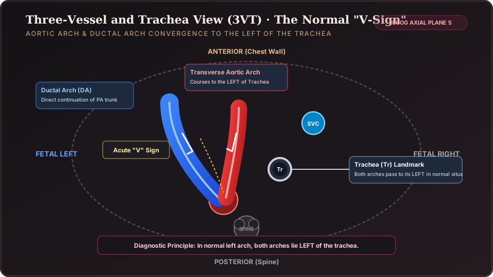 Click to zoom
Antegrade vs Retrograde Aortic Arch Flow: Ductal Steal Dynamics
Color Doppler mapping of the 3VT view. Normal heart (left): both the aortic and ductal arches show uniform forward flow (blue, away from probe). HLHS / Aortic Atresia (right): the aortic arch shows retrograde flow (red, towards the probe) due to reverse perfusion from the ductus arteriosus.
Click to zoomNormal vs Abnormal 3VV/3VT in Left-Sided Obstruction
Upper mediastinal caliber comparison across the 3VV and 3VT planes. Normal progressive caliber hierarchy PA > Ao > SVC (left) versus severe aortic hypoplasia (right) where the transverse arch is reduced to a pin-point vessel adjacent to a massive pulmonary trunk.
12. Foramen Ovale & Pulmonary Vein Doppler: Restrictive Septum in HLHS
In HLHS, the left ventricle cannot pump blood forward. Therefore, all pulmonary venous return entering the left atrium must decompress across the interatrial septum into the right atrium to be pumped out by the right ventricle.
If the foramen ovale is widely patent with an unrestrictive flap, this left-to-right decompression occurs smoothly. However, in approximately 6% to 10% of HLHS cases, the interatrial communication is severely restrictive or completely intact (premature closure).
An intact or restrictive atrial septum (RAS) in HLHS represents one of the most perilous emergencies in congenital cardiology:
- Left atrial pressure skyrockets to adult systemic levels.
- Pulmonary venous pressure rises, causing profound pulmonary venous congestion, medial arterial muscularization, and irreversible pulmonary vasculopathy in utero.
- Immediately at birth upon umbilical cord clamping, the infant experiences hyperacute profound hypoxemia, intractable acidosis, and cardiovascular collapse within minutes.
How to diagnose RAS prenatally: Do not rely on visual septal thickening alone. The gold standard is Pulsed Doppler interrogation of the pulmonary veins. In normal fetuses, pulmonary venous flow shows forward systolic (S) and diastolic (D) peaks with a minimal or absent retrograde wave during atrial systole. In restrictive atrial septum, left atrial pressure during atrial contraction forces a massive retrograde reversal velocity (A-wave reversal), with an A/B ratio >3 or high-velocity reverse flow.
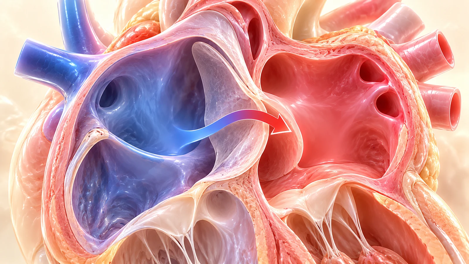 Click to zoom
Foramen Ovale Flow Direction in Left-Sided Obstruction
Interatrial septum interrogation: Normal right-to-left shunting (left) providing oxygenated umbilical blood to the left heart versus Reversed Left-to-Right shunting (right) seen in critical aortic stenosis and restrictive mitral lesions.
Click to zoomPulmonary Venous Doppler and Restrictive Atrial Septum
Pulmonary vein spectral Doppler in HLHS: Normal biphasic forward S and D waves with small forward or trivial reverse a-wave (left) versus Restrictive Atrial Septum (right) with prominent, high-velocity retrograde A-wave reversal indicating severe left atrial hypertension.
Fetuses with HLHS and a confirmed restrictive or intact atrial septum require planned delivery directly in a pediatric cardiac catheterization suite or specialized hybrid operating room with a surgical/catheterization team scrubbed and immediately ready for urgent balloon atrial septostomy or EXIT (Ex-Utero Intrapartum Treatment) decompression upon cord clamping.
13. Normal vs Abnormal Master Diagnostic Matrix
Compare the five core left-sided conditions across nine standardized clinical parameters.
| Feature | Normal Heart | Classic HLHS | Critical Aortic Stenosis | Suspected Coarctation | Unbalanced AVSD |
|---|---|---|---|---|---|
| LV Cavity & Apex | Forms cardiac apex; 1:1 symmetry with RV | Slit-like, diminutive; absent from apex | Globular, spherical; bright EFE; dysfunctional | Smaller than RV (RV>LV), but forms apex | Underdeveloped, small cavity lacking separate crux |
| Mitral Valve | Patent, biphasic inflow (A > E) | Atretic (flatline) or severely stenotic | Stenotic or secondary regurgitation jet | Patent; normal biphasic inflow velocities | Absent separate MV; common AV valve |
| Aortic Valve | Patent; laminar velocity 70–100 cm/s | Atretic plate or pin-point annulus | Thickened, doming; jet >200 cm/s or barely visible | Patent; frequently bicuspid (50–70%) | Variable; potential subaortic narrowing |
| Aortic Arch (3VT) | V-sign convergence; normal caliber | Diminutive / thread-like 1–2 mm | Normal to narrow; progressive hypoplasia | Isthmus z < -2; PA >> Ao caliber discrepancy | Usually preserved unless associated with coarctation |
| Arch Flow Direction | Antegrade (blue) | Retrograde (red) — Ductal Steal | Antegrade or bidirectional / retrograde in severe | Antegrade; diastolic flow reversal at isthmus | Antegrade |
| Interatrial Shunt | Right-to-Left (normal FO flap) | Left-to-Right decompression needed | Left-to-Right (due to high LA pressure) | Right-to-Left preserved | Large common primum ASD |
| Cardiac Crux | Preserved normal offset (TV > MV) | Normal crux topology | Normal crux topology | Normal crux topology | Effaced; single common valve |
| Key Diagnostic Clue | Apex formed by LV; 1:1 symmetry | Slit-like LV + Retrograde Arch Flow | Globular dilated LV + Bright EFE + High Jet | RV/LV > 1.3 + PA/Ao > 1.4 + Narrow Isthmus | Common AV valve committed to RV |
| Primary Uncertainty | Benign late GA dominance | Atrial septal restriction status | Pace of transition to end-stage HLHS | High false positive & negative rate prenatally | Heterotaxy & venous anomalies |
14. Prenatal Counseling & Delivery Planning: The Safety Blueprint
Counseling families facing a diagnosis of a small fetal left ventricle requires exceptional empathy, scientific transparency, and avoidance of oversimplified prognoses. The prenatal conversation must be led by a multidisciplinary team including maternal-fetal medicine subspecialists, pediatric cardiologists, neonatologists, and pediatric cardiothoracic surgeons.
Key components of the comprehensive perinatal protocol include:
Offer amniocentesis for chromosomal microarray (CMA) and gene sequencing panels. Aortic arch anomalies and IAA Type B strongly associate with 22q11.2 deletion (DiGeorge syndrome); unbalanced AVSD links with Trisomy 21 and heterotaxy genes (ZIC3, NODAL); and left heart obstruction carries familial recurrence risks (~2% to 4%). Perform detailed anatomical neurosonography and extracardiac survey.
Scan every 3 to 4 weeks (or every 1 to 2 weeks in critical AS with evolving HLHS). Monitor left ventricular growth trajectories (z-scores), development of endocardial fibroelastosis, arch flow direction, and specifically pulmonary venous flow pulsatility to detect emergent atrial septal restriction.
Delivery must be scheduled at a specialized Level IV fetal cardiac center equipped with neonatal cardiac intensive care (CICU), dedicated cardiac operating rooms, and round-the-clock catheterization capability. Spontaneous labor or routine term delivery at a community hospital carries unacceptable mortality risks for ductal-dependent lesions.
Establish immediate continuous intravenous infusion of Alprostadil (PGE1, 0.01–0.05 mcg/kg/min) upon delivery for any suspected ductal-dependent lesion (HLHS, critical AS with retrograde arch, severe arch interruption/coarctation). Keep preductal and postductal pulse oximetry continuous and conduct immediate confirmatory bedside echocardiography.
15. Twelve Practical Diagnostic Pitfalls to Avoid
Assuming Small LV Automatically Equals HLHS
Declaring HLHS without checking mitral and aortic valve patency and arch flow direction. Many small LVs represent coarctation, late gestational dominance, or loading differences.
Assuming Ventricular Disproportion Automatically Equals Coarctation
Disproportion is an indicator, not a definitive diagnosis. Over 40% of fetuses with mild late-trimester asymmetry have normal arches postnatally.
Believing a Normal 4-Chamber View Excludes Arch Coarctation
In up to 30% of coarctation cases in early mid-gestation, the four-chamber view appears deceptively symmetrical; the lesion only manifests in 3VT and arch views.
Missing Evolving HLHS in a Dilated Aortic Stenosis Heart
Mistaking a dilated, spherical LV for 'normal' size while ignoring endocardial fibroelastosis and absent forward flow. Serial scans are mandatory.
Overlooking Interatrial Septal Restriction in HLHS
Focusing solely on ventricular hypoplasia and neglecting pulmonary venous Doppler. An intact/restrictive atrial septum causes immediate fatal asphyxia at birth without emergency intervention.
Foreshortening the Ventricles by Oblique Multi-Rib Insonation
Acquiring an off-axis thoracic cut that falsely cuts off the LV apex. Always verify a true single circumferential rib prior to chamber measurement.
Failing to Interrogate the Cardiac Crux for Common AV Valve
Calling a heart HLHS when it is actually a right-dominant unbalanced AVSD. Crux effacement and a shared valve change the entire syndromic and surgical landscape.
Relying on Isolated Unvalidated Biometric Cutoffs
Using a single unindexed measurement instead of gestational-age adjusted z-scores. Cardiac dimensions must be indexed to biparietal diameter or gestational weeks.
Ignoring Persistent Left Superior Vena Cava (PLSVC)
Missing the dilated coronary sinus compressing the mitral annulus. PLSVC is a classic co-factor in left ventricular disproportion and coarctation.
Underestimating Aortic Jet Velocity by High Insonation Angle
Sampling the aortic valve with a Doppler beam >30 degrees, yielding a falsely low velocity (e.g. 1.2 m/s instead of true 2.8 m/s) and missing critical stenosis.
Over-Interpreting Mild Third-Trimester Right Dominance
Labeling a normal 34-week fetus with physiological right-sided dominance as pathological coarctation without checking arch calibers and z-scores.
Promising Parents a Definitive Prenatal Exclusion of Coarctation
Telling parents coarctation is 'ruled out'. Coarctation is inherently unpredictable prenatally; every suspect requires postnatal surveillance.
16. Ten Clinical Reasoning Scenarios & Explanations
Case Presentation: A 20-week screening scan demonstrates an RV:LV diameter ratio of 1.4:1. The mitral valve opens normally with biphasic velocity. LV forms apex. The 3VT view reveals the transverse aortic arch is smaller than the ductal arch, but color Doppler shows both arches flowing antegrade (blue).
Case Presentation: A 22-week fetus demonstrates a left ventricle that ends halfway before reaching the cardiac apex. The mitral valve is imperforate with zero color flow. The ascending aorta measures 1.2 mm. Transverse arch demonstrates pure red retrograde color Doppler.
Case Presentation: A 24-week fetus with an apparent small LV has a single common atrioventricular valve, absent crux offset, and an inlet VSD. The valve opens 70% into the RV.
Case Presentation: In a 21-week fetus, the LV is dilated, rounded like a football, and exhibits a hyper-refractile white border lining the cavity. The aortic valve leaflets are thickened with restricted opening and a peak velocity of 2.6 m/s. Interatrial flow is left-to-right.
Case Presentation: A 34-week fetus has an RV that is 15% wider than the LV. Both outflow tracts are normal, 3VT view shows symmetrical V-sign with antegrade flow, and both AV valves are patent and symmetrical.
17. OBGYNX Clinical Takeaway: LEFT IS LITTLE — FIND OUT WHY
“LEFT IS LITTLE — FIND OUT WHY”
Commit this 8-checkpoint cascade to memory. Each step evaluates a distinct physiological prerequisite for left ventricular development.
Small Left Ventricle Diagnostic Reasoning Quiz
Test your diagnostic reasoning across 10 realistic fetal echocardiography scenarios. Select the single best answer for each case. Instant rationales and comprehensive scoring are revealed upon completion.
What is the most appropriate next step in clinical decision-making?
Which congenital cardiac malformation is this finding pathognomonic for?
What is the primary diagnosis and its natural in-utero history if left untreated?
What physiological consequence does retrograde arch flow demonstrate?
Why can the prenatal diagnosis of coarctation of the aorta NOT be made with 100% certainty prior to birth?
What critical condition does this pulmonary venous Doppler profile indicate?
Which diagnostic category does this finding belong to?
What technical pitfall occurred during the initial assessment?
What is the standard neonatal management protocol pending definitive pediatric echocardiography?
Scientific References & Verified Clinical Guidelines
- Carvalho JS, et al. ISUOG Practice Guidelines (updated): fetal cardiac screening. Ultrasound Obstet Gynecol 2023; 61: 788–803. DOI: 10.1002/uog.26224.
- Moon-Grady AJ, et al. Guidelines and Recommendations for Performance of the Fetal Echocardiogram: An Update from the American Society of Echocardiography. J Am Soc Echocardiogr 2023; 36: 679–723.
- ISUOG VISUOG: Hypoplastic Left Heart Syndrome & Abnormal Four-Chamber View. International Society of Ultrasound in Obstetrics and Gynecology, 2023.
- EACTS/AEPC Guidelines Committee. Guidelines for the management of neonates and infants with hypoplastic left heart syndrome. Eur J Cardiothorac Surg 2020; 58(3): 416–499. DOI: 10.1093/ejcts/ezaa188.
- Freud PE, et al. Fetal Aortic Valvuloplasty for Evolving Hypoplastic Left Heart Syndrome: Staging and Predictors of Biventricular Outcome. Circulation 2014; 130: 638–645.
- Matsui H, et al. Prenatal diagnosis of coarctation of the aorta: predictive value of combined ventricular and great artery disproportion. Ultrasound Obstet Gynecol 2011; 38: 512–517.
- Donofrio MT, et al. Diagnosis and treatment of fetal cardiac disease: a scientific statement from the American Heart Association. Circulation 2014; 129: 2183–2242.